Study of retinal ganglion cell and retinocollicular pathway functions under pathological conditions: early mechanisms and therapeutic targets
Study of retinal ganglion cell and retinocollicular pathway functions under pathological conditions: early mechanisms and therapeutic targets
Preparation techniques for isolated rat retinal ganglion cells were established, and the electrical properties of their ion channels were investigated under brief hypoxia. The low- and high-threshold calcium currents were characterized in detail (Veselovsky M.S., Veselovska N.M., 2001).
Electrophysiological studies of retinal ganglion cells were performed using voltage- and current-clamp recordings in the whole-cell configuration on an isolated rat retina; the characteristics of high-frequency tonic firing of ganglion cells were described (Kolodin et al., 2006).
Studies of RGCs in an isolated rat retina revealed that Kv3 family voltage-gated potassium channels, which mediate rapid action potential repolarization, play a key role in generating high-frequency tonic firing of retinal ganglion cells (Grigorov et al., 2014).
Studies of RGCs in an isolated intact retina were performed under normal conditions and in experimental diabetes. The main electrophysiological parameters and spontaneous activity of rat retinal ganglion cells were examined eight weeks after streptozotocin injection; a reduction in the ability to generate high-frequency tonic action potentials and a slowing of the decay of calcium signals induced by high-frequency stimulation were observed, indicating gradual Ca²⁺ accumulation in the cytoplasm of retinal ganglion cells. These findings reflect functional alterations in ganglion cells at prepathological stages of experimental diabetes (Kuznetsov K.I. et al., 2012, 213).
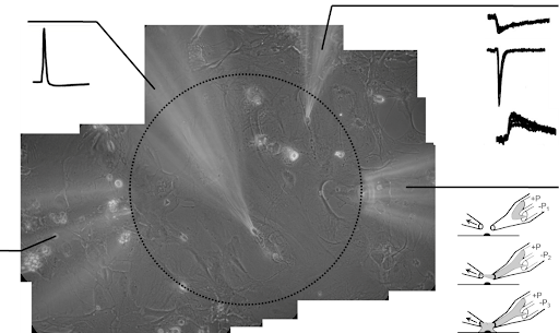
An original in vitro model of the visual retinocollicular pathway was developed—a coculture of dissociated rat retinal cells and superficial superiof colliculus (SSC) neurons. In such model, each synaptically connected pair of retinal ganglion cell-SSC neuron reflects a single fiber of the retinocollicular pathway (Patent of Ukraine for Utility Model No. 26863, 2007).
Studies of the features of neurotransmission between GCS and Superficial Superior Colliculus (SSC) neurons were conducted; functional properties of synaptic transmission between cocultured GCS and SSC neurons were described (Dumanska & Veselovsky, 2019; Dumanska et al, 2012, 2014, 2015).
The fast local superfusion technique (Veselovsky, Engert, Lux, 1996) was used to apply hypoxic solutions to synaptically connected pairs of retinal ganglion cell–SSC neuron pairs; it was shown that short-term hypoxia leads to long-term disruption of the excitatory-inhibitory balance in retinocollicular neurotransmission (Dumanska& Veselovsky, 2019, 2023).
It was demonstrated that protein kinase C signaling pathway mediates some of the hypoxia-induced effectson retinocollicular neurotransmission (Dumanska et al., 2023).
An in vitro model of hypoxia-induced dysfunction of retinal ganglion cells was developed; the effects of acute hypoxia on high-voltage-activated calcium currents in retinal ganglion cells were characterized (Dumanska et al., 2023).


