Department of Cytology
Зміст |
Science
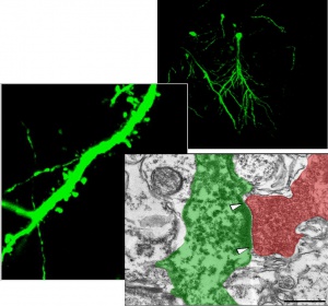
The research activity of Department of Cytology is focused on the study of molecular and cellular mechanisms of nervous tissue damages related to the development of neurodegenerative diseases (cerebral ischemia, Alzheimer's disease, Parkinson's disease) or some pathological states (perinatal pathology of the central nervous system, stress, exocrine pancreatic failure) and the investigation of approaches for prevention and correction of neurodegeneration processes. The important part of the research activity is testing of pharmacological preparations to identify their potential neuroprotective properties. Recently experiments have been started aimed at studying of possible applications of cell therapy for the treatment of brain tissue lesions caused by cerebral ischemia. In two brain ischemia models (in vitro and in vivo) it was revealed that transient ischemia induced destructive changes in neurons and remodeling of synaptic apparatus in CA1 area of hippocampus – the brain structure responsible for the formation of memory and learning.
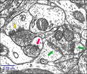
The temporal dynamics of ischemia-related delayed death of neurons in CA1 area was investigated using light and electron microscopy. The conditions applied in this study induced the delayed pyramidal cell death associated with ultrastructural modifications in excitatory synapses on dendritic spines in the stratum radiatum of the CA1 hippocampal area.The morphological ischemia-evoked remodeling of synapses manifested in following: alteration in proportion of different types of synapses, a depletion of synaptic vesicles in presynaptic terminals and changes in their spatial arrangement; a rapid increase of the postsynaptic density (PSD) thickness and length, formation of concave synapses with perforated PSD during the first 24 h after ischemic episode, a pronounced increase of the glial coverage of both pre- and postsynaptic structures.
The relationship was found between ischemia induced structural changes in neurons and activity patterns of glial cells (astrocyte hypertrophy and hyperplasia of microglia).
Oxygen-glucose deprivation (OGD) of hippocampal cultured slices revealed that hippocampal CA1 pyramidal neurons are the most sensitive to oxygen-glucose deficit, while interneurons, astrocytes and microglia are more ischemia resistant and even get activated. Study of the certain signaling pathways selective blocking reveals possible modulating effect of interneurons on viability of pyramidal neurons in hippocampus. The level of expression of subunits HIF-1α, HIF-3α, SERCA2b and PMCA1-2 in neurons of CA1 and СА3 area of hippocampus changes in response to OGD and anoxic preconditioning. The number of damaged cells in both areas of the hippocampus reduces with a stabilization of HIF-1. Developing of the original image analyzing program allowed us to determine changes in spatial patterns formed by synaptic vesicles in the CA1 synapses. It was shown that distances between individual vesicles increased while the organelles became more distant from the presynaptic membrane. It was found that during early after an ischemic damage the ratio of synaptic terminal types changed, the frequency of perforated and multiple synapses being increased. These changes may indicate ischemia-related synaptic plasticity. While researching the protective approaches against brain ischemia there was obtained the data indicating the neuroprotective properties of aromatic aminoacids, FGL - NCAM peptide mimetic, bioflavonoids, particularly korvitin, that enabled to recommend these compounds for pharmacological applications. Study of exocrine pancreatic insufficiency (EPI) in pigs discovered the dependence of morphological and functional state of hippocampal neurons on pancreas exocrine functioning. Exogenous pancreatic-like enzymes are recovered in the gut and improve growth of exocrine pancreatic insufficient pigs.
Regenerative capacity of different lines of stem cells is studied using in vivo and in vitro models of cerebral ischemia. In both models it was displayed that hippocampus-derived NPCs transplanted into the ischemic brain are able to survive for long terms after grafting, differentiate into functional mature neurons, become synaptically integrated into host circuitry. Syngeneic NPC transplantation promoted cognitive function recovery after ischemic injury in vivo.We have studied changes in proteasome proteolysis during transient occlusion of the middle cerebral artery in rats and made the comparison between changes in different types of proteasome activity and severity of ischemic injury and showed three types of decrease in proteolytic activity (trypsin-like, chymotrypsin-like, peptidyl-glutamyl peptide-hydrolyzing-like) in the brain tissues. These data suggest that ischemia may cause a severe alteration in proteasome stability and proteasome structure. Research resources of the Department are widely used for scientific practical training of students from Educational and Scientific Centre “Institute of Biology” of Taras Shevchenko National University of Kyiv, National University of Kyiv-Mohyla Academy and Kiev Branch of Moscow Institute of Physics and Technology.
Team
Stuff
- Skibo G.G. - Head of Department, MD, Prof. tel.+380 44 253-21-58, te.l./fax+380 44 256-24-42
- Kovalenko T.M. – PhD, Leading Researcher tel. + 380 44 256-24-43
- Nikonenko O.G. – PhD, Leading Researcher tel. + 380 44 256-25-70
- Tsupykov O.M – PhD, Leading Researcher tel. + 380 44 256-25-80
- Lushnikova I.V. – PhD, Senior Researcher tel. + 380 44 256 20 26
- Osadchenko I.O. – PhD, Senior Researcher tel. + 380 44 256-24-43
- Patseva М.А. – PhD, Researcher tel. + 380 44 256 24 58
- Nikandrova E.O. – Junior Researcher tel. + 380 44 256-20 26
- Savchuk O.I. – Junior Researcher tel. + 380 44 256-25-80
- Smozhanyk K.G. – Leading Engineer tel. + 380 44 256 24 45
- Mayorenko P.P. – Leading Engineer
- Melnychuk L.P. - Laboratory assistant
- Maleyeva G.
Research activities
Main areas of research
- viability and resistance of hippocampal neurons and glial cells under modeling of neuropathologies (cerebral ischemia, Alzheimer’s disease, stress); approaches for neuroprotection
- pathophysiological mechanisms of perinatal pathology of CNS and approaches for it therapeutic modulation;
- cell mechanisms of early neurodegeneration in rotenone-induced model of Parkinson’s disease;
- structural and functional peculiarities of the brain related to the gastrointestinal tract functioning;
- cell regenerative capacities for treatment of experimentally provoked brain neurodegeneration (ischemia, trauma).
Research objects
- Animals (rats, mice, gerbils, pigs)
- Cell culture (organ-like and dissociated culture)
Research methods
- modelling of neurodegeneration in vivo (cerebral ischemia, Parkinson’s disease, perinatal pathology of CNS, brain trauma, exocrine pancreatic insufficiency) and in vitro (cerebral ischemia, Alcheimer’s disease, stress);
- microscopy: light, confocal, electron;
- immunohystochemical methods;
- single-cell RT-PCR;
- immunoblotting;
- quantitative ultarstructural analysis;
- computerized image analysis;
- morphometry.
List of laboratories with which there is scientific cooperation
- Department of Cell and Organism Biology, Lund University, Sweden;
- Laboratory of Neural Stem Cell Biology&Therapy, Lund Stem Cell Center, Sweden;
- Institute of brain dynamics National Institute for Health and Medical Research (INSERM), Marseille, France;
Scientific advances. Selected publications for years 2011-2015
- Lushnikova I, Orlovsky M, Dosenko V, Maistrenko A, Skibo G. Brief anoxia preconditioning and HIF prolyl-hydroxylase inhibition enhances neuronal resistance in organotypic hippocampal slices on model of ischemic damage. Brain Res. 2011 Apr 22;1386:175-83.
- Lushnikova I, Skibo G, Muller D, Nikonenko I. Excitatory synaptic activity is associated with a rapid structural plasticity of inhibitory synapses on hippocampal CA1 pyramidal cells. Neuropharmacology. 2011 Apr;60(5):757-64.
- Tsupykov O.M., Poddubnaya A.O., Smozhanyk K.G., Kyryk V.M., Kuchuk O.V., Butenko G.M., Semenova E.A., Pivneva T.A., Skibo G.G. Integration of grafted neural progenitor cells in host hippocampal circuitry after ischemic injury// Neurophysiology. – 2011. – V.43., №.4., P. 372-375.
- Puchkov D, Leshchyns'ka I, Nikonenko AG, Schachner M, Sytnyk V. NCAM/spectrin complex disassembly results in PSD perforation and postsynaptic endocytic zone formation. Cereb Cortex. 2011 Oct;21(10):2217-32.
- Pierzynowski S, Szwiec K, Valverde Piedra JL, Gruijc D, Szymanczyk S, Swieboda P, Prykhodko O, Fedkiv O, Kruszewska D, Filip R, Botermans J, Svendsen J, Ushakova G, Kovalenko T, Osadchenko I, Goncharova K, Skibo G, Weström B. Exogenous pancreatic-like enzymes are recovered in the gut and improve growth of exocrine pancreatic insufficient pigs. J Anim Sci. 2012 Dec;90 Suppl 4:324-6.
- Woliński J, Słupecka M, Weström B, Prykhodko O, Ochniewicz P, Arciszewski M, Ekblad E, Szwiec K, Ushakova, Skibo G, Kovalenko T, Osadchenko I, Goncharova K, Botermans J, Pierzynowski S. Effect of feeding colostrum versus exogenous immunoglobulin G on gastrointestinal structure and enteric nervous system in newborn pigs. J Anim Sci. 2012 Dec;90 Suppl 4:327-30.
- Pierzynowski S, Swieboda P, Filip R, Szwiec K, Valverde Piedra JL, Gruijc D, Prykhodko O, Fedkiv O, Kruszewska D, Botermans J, Svendsen J, Skibo G, Kovalenko T, Osadchenko I, Goncharova K, Ushakova G, Weström B. Behavioral changes in response to feeding pancreatic-like enzymes to exocrine pancreatic insufficiency pigs. J Anim Sci. 2012 Dec;90 Suppl 4:439-41..
- Никоненко А.Г. Введение в количественную гистологию. Киев: Книга плюс, 2013, 256с. 500 прим. 15,5 друк. арк.
- Rybachuk О. А, Levin R. E., Кyryk V. М., Susarova D. K., Tsupykov О. M., Smozhanik E. G., Butenko G. M., Skibo G. G., Troshin P. A., Pivneva Т. А. Effect of a water soluble derivative of fullerene C60 on the features neural progenitor cells in vitro, Cell and organ transplantology, 2013, 1, № 1, 96-107
- Nikonenko I, Nikonenko A, Mendez P, Michurina TV, Enikolopov G, Muller D. Nitric oxide mediates local activity-dependent excitatory synapse development. Proc Natl Acad Sci U S A. 2013, 110(44):E4142-4151.
- Pierzynowski S, Ushakova G, Kovalenko T, Osadchenko I, Goncharova K, Gustavsson P, Prykhodko O, Wolinski J, Slupecka M, Ochniewicz P, Weström B, Skibo G. Impact of colostrum and plasma immunoglobulin intake on hippocampus structure during early postnatal development in pigs. Int J Dev Neurosci. 2014 Mar 15. pii: S0736-5748(14)00037-9
- Rybachuk O. A., Kyryk V. M, Poberezhnyi P. A., Butenko G. M., Skibo G. G., Pivneva T. A. Effect of bone marrow multipotent mesenchymal stromal cells on the neural tissue after ischemic injury in vitro, Cell and organ transplantology, - 2014, 2, № 1, p. 96-100.
- Tsupykov O, Kyryk V, Smozhanik E, Rybachuk O, Butenko G, Pivneva T, Skibo G. Long-term fate of grafted hippocampal neural progenitor cells following ischemic injury. J Neurosci Res. 2014 Aug;92(8):964-74.
- Voytenko LP, Lushnikova IV, Savotchenko AV, Isaeva EV, Skok MV, Lykhmus OY, Patseva MA, Skibo GG. Hippocampal GABAergic interneurons coexpressing alpha7-nicotinic receptors and connexin-36 are able to improve neuronal viability under oxygen-glucose deprivation. Brain Res. 2015 Aug 7;1616:134-45.
- Goncharova K, Skibo G, Kovalenko T, Osadchenko I, Ushakova G, Vovchanskii M, Pierzynowski SG. Diet-induced changes in brain structure and behavior in old gerbils. Nutr Diabetes. 2015 Jun 15;5:e163.
- Tsupykov O. Ultrastructural analysis of murine hippocampal neural progenitor cells in culture. Microsc Res Tech. 2015 Feb;78(2):128-33.
- Скибо Г.Г., Коваленко Т.М., Лушнікова І.В., Півнева Т.А., Осадченко І.О., Цупиков О.М. «Експериментальна ішемія мозку», Київ: Наукова думка, 20 др.ар.


